
Author: Service Lifter

Orthodontic Headgear

Sometimes, braces alone aren’t enough to move teeth into a better position, or to correct trouble with the bite or remedy problems in the growth of the jaws. In those situations, special appliances may be recommended. Orthodontic headgear is the general name for an appliance, worn partly outside the mouth, which creates just enough force to move the teeth properly and guide the growth of the face and jaws.

There are several different types of orthodontic headgear, each designed to work best in a specific situation. A treatment program will be designed to address your individual needs, and select the most appropriate type of headgear; you will also be instructed on its use and care. It’s important for you to follow instructions carefully so that you can achieve the best results from your treatment.

Types of Headgear

One common type of headgear is called the cervical pull type. This appliance has a U- shaped wire that attaches to the bands on your back teeth, and a strap that is worn behind your neck. A similar device is the high-pull headgear, which also has a wire connecting to the teeth, plus a strap that goes behind and over the head.

Overjet and Underbite.

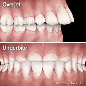
These types of headgear are typically used to correct an excessive horizontal overbite (technically called an “overjet”) in children, by holding back the growth of the upper jaw. They can also be used for adults who need help maintaining a proper bite and correct tooth spacing after tooth extraction. Generally, these types of headgear are designed to be worn from 12-14 hours each day.

Another type of headgear is the reverse-pull or “facemask” type. This appliance is generally used to correct an underbite. It gently pulls the upper jaw forward (instead of back), which allows it to catch up with the lower jaw. It consists of two pads — one resting on the forehead, the other on the chin — connected by a vertical frame. Elastics or wires, which connect from the frame to the braces, exert the pulling force. It may be necessary to wear this appliance from 14-16 hours daily.

Making Headgear Work Depends on You

Whichever type of headgear you’re wearing, there are some important things you should know. Probably the most essential one is this: In order for it to be effective, you must carefully follow instructions about wearing your headgear — that means putting it on each day for the time specified. If you wear headgear at night and you miss one night, you must make up the time the following day — otherwise, everything you’ve accomplished in the previous seven days of wear could be wiped out!

It’s normal to feel some discomfort as you get used to wearing orthodontic headgear. Fortunately, if you wear it faithfully, the discomfort generally goes away in a few days. An over-the-counter pain reliever like Ibuprofen, and/or a soft diet, may be recommended to help you adjust.

From time to time you may also experience some soreness when chewing, or even a little looseness in the first molars. This is normal, and it shows the appliance is working. However, if you have unusual pain, notice that the anchor band on your first molar (the one the headgear attaches to) has come loose, or find that the headgear suddenly seems not to fit correctly, it could signal a problem that requires immediate attention.

Maintain Your Headgear — And Your Oral Health

To keep your orthodontic headgear working as it should — and to maintain your overall oral health — it’s important that you follow all instructions about care and cleaning. It’s also important that you learn to put headgear on and take it off properly and safely. Remember to bring it with you every time you have an orthodontic appointment — but leave it behind when you’re playing sports, or even horsing around in the living room!

Wearing orthodontic headgear may seem like a big adjustment — and nobody would deny that it takes some getting used to. When everyone works together, it’s possible to achieve your goal: a beautiful smile that you’ll have for your whole lifetime.

Digital X-Rays

It’s almost impossible to imagine the practice of dentistry without x-ray technology. Radiographs (x-ray pictures) allow dentists to diagnose and treat problems not yet visible to the naked eye, including early tooth decay, gum disease, abscesses and abnormal growths. There is no question that since x-rays first became available a century ago, this diagnostic tool has prevented untold suffering and saved countless teeth. Now, state-of-the-art digital x-rays have made the technology even safer and more beneficial.

Digital x-ray technology uses a small electronic sensor placed in the mouth to capture an image, which can be called up instantly on a computer screen. When digital x-rays first became available about 20 years ago, they immediately offered a host of advantages over traditional x-ray films, which require chemical processing. Most importantly, they cut the amount of radiation exposure to the dental patient by as much as 90%. While faster x-ray films have been developed over the years that require less exposure, making that difference less dramatic, a digital x-ray still offers the lowest radiation dose possible.

Advantages of Digital X-Rays

Besides minimizing radiation exposure, digital x-rays offer numerous advantages to dentists and patients alike. These include:

  • No chemical processing & no waiting. Because there is no film to process with digital x-rays, there is no waiting for pictures to develop — and no toxic chemicals to dispose of. Your dentist can immediately show you the pictures on a computer screen for easy viewing.
  • A clearer picture. It’s possible to get more information from digital x-rays because they are sharper and can be enhanced in a number of ways. The contrast can be increased or decreased, and areas of concern can be magnified. It’s even possible to compare them on-screen to your previous x-rays, making even the minutest changes to your tooth structure easier to detect.
  • Easy sharing and storage. Digital x-rays provide a better visual aide for you, the patient, to understand your diagnosis and treatment options. They can be e-mailed to different locations; they are also far less likely to be misplaced.

X-Rays and Your Safety

While digital technology has minimized the health risks of x-rays, it has not entirely eliminated it. X-rays are a type of radiation used to penetrate the tissues of the body to create an image. In doing so, there is always a slight possibility of causing changes at the cellular level that might lead to future disease. Of course, there are sources of radiation present in the daily environment — the sun, for example — that can also cause disease. It’s important to note that the chance of this happening is thought to be cumulative and not based on a single exposure. Still, x-rays are not considered risk-free regardless of how technology reduces your exposure. That’s why dentists will only use them when the benefit of obtaining better diagnostic information outweighs the procedure’s small risk. This is particularly true of computed tomography or CT scans, which can raise the level of exposure, yet yield a tremendous amount of information per scan. No matter which technology is being used, each case is considered individually, and your safety is always paramount. If you have questions about why an x-ray is being recommended for you, please feel free to ask.

Corrective Jaw (Orthognathic) Surgery

In many cases, problems with the bite or the alignment of the teeth can be corrected using today’s advanced methods of dental and orthodontic treatment. Sometimes, however, skeletal and dental irregularities aren’t so easy to remedy, even with the most up-to-date non-surgical techniques. That’s when corrective jaw surgery (also called orthognathic surgery) may be recommended.

While it may sound like a complex and demanding treatment, jaw surgery isn’t always so serious. It’s often performed as a routine in-office procedure — for example, to extract impacted wisdom teeth (molars that don’t fully protrude through the gums), or to place dental implants in the jaw. However, it can also be used to remedy severe orthodontic problems involving the relationship between the teeth and jaws, including the correction of underbites (the most frequent surgical correction) and congenital abnormalities (birth defects) related to jaw development. It can even help alleviate sleep apnea, a potentially life-threatening condition.

Who Can Benefit From Jaw (Orthognathic) Surgery

People who have problems related to the jaws, tooth alignment and facial asymmetries, which create difficulties chewing, talking, sleeping, or carrying on routine activities, may benefit from having orthognathic (jaw) surgery. These procedures can also be used to correct aesthetic issues, such as a protruding jaw, a congenital defect, or an unbalanced facial appearance.

After a thorough examination, it can be determined if you’re a candidate for orthognathic surgery. In general, if orthodontic treatment can solve the problem, that’s where you will start. Yet, while orthodontics can successfully align the teeth, it’s sometimes the jaws themselves that need to be brought into line. In most cases, orthodontic appliances, such as braces and retainers, will be used before and after the surgical phase of treatment, to ensure that you end up with an effective — and aesthetically pleasing — result.

Conditions that can be successfully treated with corrective orthognathic surgery include the following:

  • Open bite, protruding jaw or receding chin
  • Congenital defects such as a cleft palate
  • Malocclusions (bite problems) resulting from underbites or severe overbites
  • Obstructive Sleep Apnea, when more conservative methods fail
  • Difficulty swallowing, chewing, or biting food
  • Chronic jaw or jaw joint (TMJ) pain and headache
  • Unbalanced facial appearance from the front or side
  • Inability to make the lips meet without straining
  • Chronic mouth breathing and dry mouth
  • Facial trauma

The Surgical Procedure

While every patient’s needs are different, it’s possible to outline some typical steps in the process. The first (and perhaps most important) component is consultation and planning among members of the dental team, including the restorative or general dentist, the orthodontist, and the oral surgeon. Your overall plan may involve orthodontic treatments along with surgical procedures. Using diagnostic images, 3-D models and advanced software, a step-by-step plan will be developed for the entire process. It’s even possible in many cases to show you a picture of what you’ll look like when your treatment is finished.

The surgery itself may occur in a hospital or an office setting, with the type of anesthesia that’s most appropriate for the procedure, and for your comfort. Because the actual surgery is generally performed inside the mouth, it often leaves no visible traces. After the procedure, minor pain and swelling can be controlled by over-the-counter or prescription pain medication. A soft or liquid diet may be recommended for a period of time following the procedure.

Following surgery, your condition will be closely monitored — as will your progress through each phase of the treatment plan. When it’s complete, you can enjoy the benefits of improved functionality and an enhanced appearance.

Missing Teeth

If you are missing one or more of your adult teeth, you’re not alone — an estimated 178 million Americans have the same condition. Many try to get along without all of their teeth, and suffer from some of the familiar problems that go with the territory: smiles that don’t look as appealing as they once did, problems eating certain foods, and awkwardness or embarrassment in social situations.

Yet there are other problems associated with tooth loss that are less obvious, but could have more of an impact on your health. These include nutritional difficulties, oral health issues, and bone loss.

Nutritional Difficulties

It is well established that eating plenty of fruits and vegetables and avoiding highly processed foods are essential parts of good nutrition. But many foods become difficult to chew if you have missing teeth — and those “challenging” foods are often the same ones that offer the greatest nutritional value. Softer foods are easier to eat, but they are often highly processed, and offer little nutritional value. Along with the possibility of malnourishment, a poor diet can lead to a compromised immune system and a decline in overall health.

Oral Health Issues

If you have only one or two missing teeth, you may not feel an urgent need to replace them now. Yet the problems that stem from missing teeth don’t get better — and in time, they can get much worse. Teeth aren’t fixed solidly in the jaw; instead, when even one tooth is missing, the remaining teeth tend to “drift” into new positions. This can cause a cascade of oral health problems, including unstable tooth positions, excessive tooth wear, bite problems, a greater chance of developing tooth decay and periodontal disease — and the loss of even more teeth.

Bone Loss

Consequences of Tooth Loss.You can’t see the bone inside your jaw — but the consequences of bone loss are very real. Bone tissue needs stimulation to maintain its volume and density. When teeth are lost, the jaw bone that once supported them loses stimulation and begins to deteriorate. It can lose one-quarter of its width in just one year, and even more as time passes. As the jaw becomes smaller, facial height (the distance from nose to chin) decreases, and facial features lose support. The chin rotates forward, the corners of the mouth turn downward (as if frowning), and the cheeks can appear hollow. Loss of bone in the jaw can give you the appearance of being many years older than you actually are.

The consequences of tooth loss are very real, even if they are less visible than a gap in your smile. That’s why it is so important not to put off the replacement of missing teeth.

Loose Teeth & Bite Problems

When you are a child, your first loose tooth can be cause for celebration; when you are an adult, it definitely is not. Biting and chewing with a tooth that is not securely attached in its socket can be difficult or even painful — and any tooth that becomes loose is at risk of needing to be removed or, at worst, falling out. It’s often possible to prevent that from happening, but quick action is required.

Causes

The most common reason for tooth looseness is periodontal disease — a bacterial infection of the gum and/or bone tissues that surround and support your teeth. The infection results from bacterial plaque that sits on your teeth in the absence of effective oral hygiene. Over time, periodontal disease will cause gum tissue to detach from the teeth as plaque and tartar formation increases and tooth-supporting bone is lost. As more bone is lost, teeth gradually become loose and are unable to withstand normal biting forces. If severe periodontal disease remains untreated, loose teeth will eventually fall out.

Another common contributor to the loosening of teeth is a clenching or grinding habit that generates too much biting force. This force can stretch the periodontal ligaments that join the teeth to the supporting bone, making your teeth looser. These habits can accelerate bone loss and also cause jaw pain and excessive tooth wear.

Treatment

Just as the causes of loose teeth can be biological (disease) or mechanical (too much force) — or both — so, too, are the treatments. Let’s first take a look at the biological approach.

To control gum disease, a dental professional must thoroughly clean the teeth of plaque and harder deposits (tartar or calculus) in which bacteria thrive; this includes the tooth-root surfaces beneath the gum line. At the same visit, you will be instructed on effective oral hygiene techniques and products to use at home. Often this type of deep cleaning, combined with improved oral hygiene, will reduce inflammation and heal the gums enough to cause some tightening of the teeth.

The mechanical approach involves modifying the forces that are applied to the teeth. This can be achieved in a number of ways. For example, an occlusal (bite) adjustment can be performed by carefully reshaping minute amounts of tooth surface enamel. This changes the way upper and lower teeth contact each other, redirecting and lessening the force. Replacing broken fillings and restoring worn teeth is also sometimes needed to create a more balanced bite, even when teeth are not loose.

Splinting teeth.
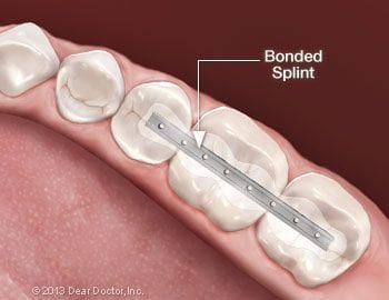
It is also possible to reduce stresses on teeth by temporarily or permanently splinting them together like fence pickets so that any biting force is distributed among groups of teeth rather than individual loosened teeth. The splint is a very small metal bracket bonded to the backs of or tops of the teeth.

If clenching or grinding habits are a problem, a custom-made bite guard (also called an occlusal splint) can be worn when needed. Placed in the mouth at night or in times of stress, it can protect the teeth from the consequences of too much biting force. This can also be helpful in preventing excessive tooth wear, and sometimes in relieving jaw pain.

Loose teeth can be successfully treated with both biological and mechanical techniques. A thorough examination will be needed to determine the best approach. So if you are experiencing tooth looseness, don’t wait — the sooner this problem is addressed, the more likely you are to keep your natural teeth.

Cone Beam CT Imaging

In the early 20th Century, not long after X-rays were discovered, medical professionals recognized their value as diagnostic tools: They could clearly reveal structures hidden inside the body without the need for risky surgery. At the dawn of the 21st century, a revolutionary new technology has entered the diagnostic arena. Today, Cone Beam Computed Tomography (CBCT) promises to change the way many dental problems are diagnosed and treated.

Cone Beam CT has some similarities with conventional X-rays, and also with the standard CT scans you would get in a hospital setting. But it’s a quantum leap forward in technology and diagnostic precision. For the dentist, it offers the ability to visualize intricate structures inside the mouth, such as root canals, nerves and sinuses (air-filled spaces) in the jaw — in three dimensions — without surgery. For the patient, it can reduce the need for invasive procedures, shorten treatment time and offer the chance for a better outcome.

The detailed diagnostic images that CBCT provides have made it an essential tool in many dental specialties. But, as with any diagnostic tool that uses radiation, the medical benefits offered must be weighed against the (small) potential risks of the procedure.

How Cone Beam CT Works

X-rays, like visible light, are a form of energy on the electromagnetic spectrum. Just as light makes an image on photographic film (or a digital camera sensor), X-rays can also form an image. The difference is that energetic X-rays can penetrate bone and soft tissue, and reveal its hidden structure by their varying degrees of absorption; in other words, they form a grayscale picture of what’s underneath the surface. But conventional X-rays are limited: Like a still-life picture, they show only one perspective on the scene.

Now imagine a “flip book” — the kind of small book made up of a series of pictures, each slightly different. When you rapidly page through it, you may see (for example) an animated cartoon or a still subject from different perspectives. If you could put together a flip book made from a series of X-ray “slices” of the same subject, taken at slightly different angles, you would be able to create an “animation” of the X-rays. And from there, it’s only one more step to making a 3-D model.

That’s exactly what CBCT scanners do. Using a rotating imaging device that moves around the patient’s head, the scanner records between 150 and 600 different X-ray views in under a minute. Then, a powerful computer processes the information and creates a virtual model of the area under study. When it’s done, the model appears as a three-dimensional image on a computer screen: It can be rotated from side to side or up and down, examined in greater or less detail, and manipulated in any number of ways — all without the patient feeling any discomfort… or even being present.

Where Cone Beam CT Is Used

The ability to see fine anatomical structures in 3-D has proven invaluable in treating conditions in many areas of dentistry.

  • Orthodontics: Having accurate information on the position of teeth and jaws helps determine exactly how and where teeth should be moved.
  • Dental implants: Detailed CBCT images are used to determine the optimum location for the titanium implants while avoiding nerves, sinuses and areas of low bone density.
  • Orthognathic Jaw Surgery and Temporo-mandibular Joint (TMJ) Disease: Patients benefit when the specialists who treat these conditions can evaluate their anatomy with the three-dimensional perspective that cone beam CT provides.
  • Oral Surgery: Treatment for tumors or impacted teeth is aided by the level of fine detail shown in these scans.
  • Endodontics: Dentists performing intricate procedures (like complex root canals, for example) can benefit from a clearer visualization of the tooth’s anatomy.
  • Sleep Apnea: Imaging the tissues and structures of the nose, mouth and throat can aid in diagnosis and treatment of this dangerous condition.

Could Cone Beam CT Benefit You?

Each patient’s situation is different, and must be carefully considered by a clinical professional before any test or procedure is performed. While CBCT delivers a smaller dose of radiation (X-rays) than many other diagnostic tests, it still carries a small risk — particularly for younger patients, or those with other health problems. As is the case for any medical procedure, all risks, benefits and alternatives are taken into account before the procedure is recommended.

Digital Dental Impressions

For years, whenever you needed a dental crown (cap), your dentist had to make molds of your teeth which required taking an impression of your teeth. A tray filled with a goopy, putty-like material was used so that a three-dimensional model of the prepared tooth could be created. Using this mold, a dental lab could custom-craft the new crown.

However, as we journey further into the technology-driven 21st century, this traditional methodology is being replaced with virtual models — made using small, handheld “wands” that employ a digital camera and some reflective dust.

Here’s how it works

The initial phase of restoration, preparing the tooth surface, remains virtually the same. First, any dental decay must be removed, and the remaining tooth must be shaped so that a crown or filling can be fitted properly. This will allow the tooth to be restored to its original shape, look, and function. Next, the area is lightly dusted with a reflective material (not a goopy impression material) so that multiple images of your tooth’s surface can be recorded with a small scanning wand. Later, the computer component is connected to the scanning wand and these separate images are combined into a computer-generated 3D image.

Digital Dental ImpressionsThis remarkable tool uses blue wavelength light to precisely capture the unique nooks and crannies of your tooth’s surface and make a highly accurate 3D digital model. It makes it possible to instantaneously examine your tooth, and your bite. It’s possible to identify any additional prep work required for new crowns, veneers and fillings right then and there; to implement any needed changes; and to rescan the tooth to create a new series of images and 3D model.

Once the image capture and prep work are satisfactory, your images are sent on to the lab for fabrication. This technique makes it possible to create a crown or a filling that can often be completed during a single office visit.

How this technology benefits you

  • Finally, you can say goodbye to the goop, gagging, discomfort, and anxiety you’ve experienced in the past with traditional dental impression materials!
  • It enables the immediate assessment of whether or not your tooth has been properly prepared for restoration.
  • This technology is ideal for fabricating restorations such as new crowns, veneers and fillings for teeth — often possible in one office visit.
  • It takes less time than traditional dental impressions.

Fixed Dentures

If you have lost an entire arch of teeth (top and/or bottom), or are soon to have your remaining teeth removed because they are too unhealthy to save, you may be able to replace them with fixed dentures supported by dental implants. Doctors and patients alike prefer fixed over removable dentures because they:

  • Look, feel and function just like natural teeth
  • Don’t slip when you eat or talk
  • Prevent bone loss in the jaw
  • Last a lifetime

How It Works

Dental implants serve the same purpose as the roots of natural teeth: anchoring the replacement teeth to your jawbone. Just like natural tooth roots, they lie under the gum line and therefore are not visible in the mouth. Only the lifelike prosthetic teeth attached to them (the fixed denture) can be seen by you or anyone else. Because dental implants are made of titanium, a metal that has the unique ability to fuse to living bone, they are extremely stable and reliable. How many implants are needed? The number varies because each individual has unique conditions: Depending on the volume and density of the bone in your jaw, you will need as few as four implants or as many as six for your new teeth to function as well as a set of healthy, natural teeth.

What to Expect

Implant and Replacement TeethThe surgery to place dental implants that support a fixed denture is a simple, routine procedure carried out in an office setting, under local anesthesia in most cases. (If you need to have failing teeth removed, that will be done first, often the same day your implants are placed). After numbing the area, the appropriate number of implants will be placed in your jaw at precisely planned angles and positions to maximize support and avoid anatomical structures such as nerves and sinuses. Depending on how many implants are needed, the surgery can take anywhere from one to three hours. Most people who have dental implants placed find that any post-operative discomfort can be managed with over-the-counter anti-inflammatory medication such as ibuprofen or acetaminophen. Some don’t even need to take that.

What happens immediately after surgery will depend on what’s best to promote healing in your individual situation. Sometimes a set of temporary teeth can be attached immediately, so that you can leave the office with new teeth. A few months later, your permanent replacement teeth with be installed. In other cases, the implants will be left to heal for several months before any teeth are attached. Sometimes that is the best way to insure that the implants remain undisturbed as they go through the process of fusing to your jawbone, which is known as osseointegration.

In either case, you will need to go easy on your newly placed implants during the crucial healing phase following surgery. You will be advised to eat a softer diet and avoid hard, chewy foods until the process of osseointegration is complete — about three months. While this may seem like a long time, keep in mind that people who wear removable dentures often avoid these foods permanently. The good news is that once your implants have fused to your jawbone and your new permanent teeth are attached, you will be able to eat anything you want. In fact, you are likely to forget you even have dental implants!

Antibiotic Premedicationfor Dental Treatments

Antibiotics are widely prescribed to control bacterial infections. Sometimes they are given before a medical or dental procedure, to prevent a possible infection from occurring; this practice is called “antibiotic prophylaxis.” In the recent past, physicians and dentists advised that people with certain medical conditions – including a number of heart problems and several types of bone or joint replacements – should always take antibiotics before many routine dental procedures. Today, their advice may be different.

A growing body of evidence now indicates that far fewer patients need to take this preventive step than was previously thought. As a result, the guidelines for prescribing antibiotic prophylaxis have recently changed – and they may do so again in the future. Why are the recommendations changing – and what do you need to know about taking antibiotics before coming to the dental office?

The Risk of Infection

We all know that bacteria – both helpful and harmful types – thrive in many parts of the body, including the mouth. Whenever circumstances make it possible for these microorganisms to enter the bloodstream, there’s a slight risk that a bacterial infection may develop. This could occur in many dental procedures – and it could also occur during routine activities like chewing, brushing and flossing. In most cases, the risk is so small that the chance of a having bad reaction to antibiotics (while rare) is far greater than the chance of developing an infection; therefore, antibiotics aren’t routinely used.

Some people, however, need to take extra precautions before having dental procedures. If you have been treated for some types of heart disease, or have had certain orthopedic procedures (including total joint replacement), we may advise taking antibiotics to protect against even a remote chance of infection. Recommendations are made on an individual basis, taking into account your medical history and a clinician’s healthcare experience.

Guidelines for Antibiotic Premedication

Prophylactic antibiotics might be recommended before dental procedures if you have one or more of the following heart conditions:

  • A heart transplant
  • Artificial heart valves
  • A history of infective endocarditis
  • Some types of congenital heart problems – particularly if they haven’t been completely repaired, or if their treatment involves prosthetic material

If you have undergone a joint replacement procedure, prophylactic antibiotics might be recommended if you also have one or more of the following risk factors:

  • A systemic inflammatory disease such as rheumatoid arthritis or lupus erythematosis
  • A weakened immune system resulting from HIV, cancer, radiation or chemotherapy, or another cause
  • Insulin-dependent (type I) diabetes or hemophilia
  • A history of previous infection in a prosthetic joint
  • Undernourishment or malnourishment

There are other circumstances where taking prophylactic antibiotics would be a prudent step; there are also a number of situations where these medications might have been recommended in the past, but aren’t currently required in all cases. For example, the presence of a benign heart murmur, a pacemaker or defibrillator, and certain heart diseases or congenital defects don’t automatically mean that antibiotic prophylaxis will be needed.

In recent years, reports of drug-resistant bacteria and harmful side effects from some medications have increased public awareness of the consequences of overusing antibiotics. Fortunately, new scientific research is helping healthcare professionals make better, evidence-based treatment decisions on antibiotic use. If you have questions about whether you should take antibiotics before dental procedure, don’t hesitate to ask.

Adolescent Orthodontic Care

For many teens, braces are a rite of passage: They’re one more example of the changes adolescents go through at this time — along with growth in stature, edgier tastes in clothes and music, and an increasing degree of self-awareness. But is there any particular reason why orthodontic appliances and teenagers seem to go together? In a word: Yes.

There are several good reasons why adolescence is the optimal time for orthodontic treatment, though occasionally even earlier intervention is called for. One has to do with the development of the teeth: There’s no set timetable for every kid, but generally by the age of 11-13 the deciduous (baby) teeth have all been lost, and the permanent ones have largely come in. This is the time when we can go to work correcting the problems that cause a bad bite (malocclusion), improper tooth spacing or poor alignment.

Orthodontic problems don’t improve with age — they simply become harder to treat. It’s easier to treat many orthodontic problems during adolescence because the body is still growing rapidly at this time. Whether standard braces are used, or appliances like palatal expanders, improved appearance and function can be created in a short period of time. In later years, when the bones of the face and jaw are fully developed, many conditions become more difficult (and costly) to treat.

There’s even a social element to getting orthodontic treatment in adolescence. If you need braces, you’re not alone! Chances are you’ll see some of your classmates in the dental office, and you may even make new friends as you go through the process together. When it’s done, you’ll have a smile that you can really be proud of, and benefits that will last your whole life.

The Orthodontic Treatment Process

What can you expect when you have orthodontic treatment? It all depends on what kind of treatment you need. At your first appointment, pictures and radiographic (X-ray) images of your mouth are usually taken, along with impressions of your teeth, so that a model of your bite can be made. This information will be used to develop a treatment plan. It may involve regular braces, with or without elastics (rubber bands). A specialized appliance may also be recommended for a period of time. Here are some of the most commonly used orthodontic appliances:

Metal braces.
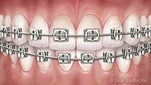
Metal Braces need no introduction. But you might be surprised to find they’re smaller and lighter than ever. They may even offer some customized options, like colored elastic ties on the brackets.

Clear braces.
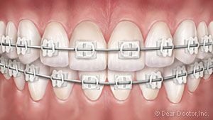
Clear Braces feature brackets made of ceramic or composite materials which blend in with your teeth, making them harder to notice. They’re suitable in many situations, but they cost a little more.

Clear aligners.
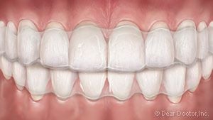
Clear Aligners for teens is a series of removable, clear plastic trays that gradually straighten teeth as they’re worn (for 22 hours per day). Formerly recommended only for adult patients, they now come with special features — like compliance indicators to tell how often you’ve been wearing them — that make them appropriate for teens in some situations. The advantage: they’re practically invisible!

Lingual braces.
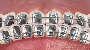
Lingual Braces offer the most unnoticeable form of orthodontic treatment because they are attached to the back (tongue side) of the teeth, where they cannot be seen at all.

Other orthodontic appliances may be recommended in some cases, where major tooth or jaw movement is needed. They can range from small devices that fit inside the mouth to external headgear. But don’t worry: You’ll get used to them, and they’re temporary — but they provide a long-term benefit in a short time.

How Long Will I Wear Them?

There’s no one answer that fits everyone: It all depends on what has to be done in your individual situation. Generally, however, the active stage of orthodontic treatment lasts 6-30 months. Afterwards, you will wear a retainer for another period of months. When your orthodontic treatment is complete, a new smile will be yours for a lifetime.