
Author: Service Lifter

Gum Grafting

If gum recession is affecting your health or your appearance, a procedure called gingival (gum) grafting may be recommended. Although it might sound scary, a variety of gum grafting procedures are routinely performed by periodontists (specialists in the area of gingival tissue), and by some general dentists with specialized training in this field.

Gum grafting involves carefully placing a small amount of new tissue in an area where little or no gum tissue currently exists — typically recommended to prevent further gum recession or to cover root surfaces of your teeth that have become exposed. The tissue used in this procedure may come from a variety of sources but usually is taken from the palate (roof of the mouth), after the area has been numbed for your comfort. Then it is delicately sutured (stitched) in place where it’s needed, using suturing material which may be finer than a human hair.

Your body’s natural recovery process takes over after the grafting procedure is complete. During this time, new blood vessels grow into the graft and help it to become integrated with the surrounding tissue. A successful graft can reduce or eliminate problems like tooth sensitivity and further gum recession, as well as improve the aesthetics of your smile.

How do you know if you need a gum graft? Often, you can clearly see or feel the problem. Exposed tooth roots can make your teeth look overly “long,” and they also generally appear somewhat darker than your pearly white crowns. Gum recession is a condition that can increase with age, as we recognize when we hear the phrase “long in the tooth.” But the potential problems of gum recession aren’t just cosmetic — they can also seriously impact your oral health.

A Closer Look at Your Gums

Inside your mouth, gum tissue forms a barrier that resists the vigorous mechanical (and microbial) effects of eating, chewing and biting. Gums may begin to recede, or shrink down, for several reasons.

One is ineffective oral hygiene: This includes both inadequate brushing, which fails to remove dental plaque; and excessive brushing, which erodes the tissue. Foreign objects in the mouth that rub against the gums, like poorly fitting removable dentures, tongue bolts, or even fingernails, can also contribute to gum recession.

When the tooth’s roots lose the protection of healthy gum tissue, they can become extremely sensitive to hot or cold temperatures, causing pain when you consume many foods and drinks. Worse, missing gum tissue makes it easier for bacteria to penetrate the roots and cause decay — or for minor trauma to result in rapid gum recession. In either case, the eventual result may be tooth loss. That’s why proper treatment of gum recession is so important.

Treating Gum Recession With Gingival Grafting

After a thorough examination and assessment of your teeth, gums and overall health, the gingival grafting procedure may be recommended. The tissue used in this procedure may be obtained from a variety of sources: an area next to the area of recession, from the hard palate, or donated tissue from another person, which has been medically processed to make it safe to use.

There are several different methods of grafting. For example, if you have lost gum tissue in an area of your mouth that’s not highly visible when you talk or smile, additional gum tissue can be placed there to prevent further recession without trying to cover the tooth-root surface completely. This procedure is referred to as a free gingival graft (since the tissue is separated from its blood supply), and it takes about 45 minutes to perform. It is very predictable with a very high success rate. If the objective is to cover root surfaces, the procedure becomes more complicated and will take longer, but is still very successful.

Grafting procedures are typically carried out under local anesthesia, and are generally pain-free. You should experience very little discomfort after the surgery, even if tissue has been removed from your palate, because that donor area is usually covered by a soothing, liquid band-aid-like material. Fortunately, any discomfort that you do have can be alleviated with over-the-counter or prescription anti-inflammatory medication, and it is generally short-lived. A soft diet (and an antibiotic) may also be recommended for a week or so thereafter, enabling the tissues to heal fully; otherwise, your normal activities will not be limited.

Keeping Your Gums Healthy

Following a gum graft, it’s more important than ever to practice good oral hygiene; this will help prevent gum problems from developing in the future. Your regimen should include proper brushing with fluoride toothpaste, daily flossing, a healthy diet and regular dental and periodontal checkups. Maintaining healthy gums is a key to keeping up your overall oral health — and preserving your smile.

Orthodontics & Dental Hygiene

You already know that maintaining good oral hygiene is important for everyone — but when you’re having orthodontic treatment, it’s even more critical. Why? Because, while the appliances (such as braces or clear aligners) you may need to wear during treatment are very effective in correcting misaligned teeth, they can also trap food particles easily. Keeping your teeth (and your appliances) clean is a little harder — but you can do it! Here’s a look at why good oral hygiene is so important during orthodontic treatment, and some tips on how you can keep it up.

The major enemy of oral health is plaque. Food that becomes trapped near tooth surfaces can lead to the formation of plaque — a thin coating of microorganisms and organic debris (biofilm) containing potentially harmful bacteria. Braces or other appliances make it harder to remove plaque. The bacteria in plaque digest the sugars in food, producing acids which may erode teeth and irritate gums. This can cause cavities, white spots on teeth, gum disease and bad breath.

Keeping plaque under control is one of the most effective means of maintaining strong, healthy teeth and gums. There are three general ways to do it: through diet, daily maintenance, and regular professional care. Taken all together, they’re your teeth’s best defense.

Diet and Decay

Controlling your diet involves avoiding foods that could increase your risk of developing tooth decay. That means cutting down or eliminating foods with an excess of sugar, like soda, sweets, and ice cream. It also means avoiding foods that could easily become stuck in your braces, like toffee, gum, licorice and caramels.

Foods that are very hard or extremely sticky can also cause physical damage to orthodontic appliances. Certainly braces or retainers with broken wires or loose brackets aren’t working to straighten your teeth! You should avoid foods like hard candies or nuts, beef jerky and hard pizza crust. Keep eating healthy foods like carrots and apples — but cut them into bite-sized pieces first! And don’t chew on ice, pencils, or your nails: these habits can cause damage to your appliances, and even result in chipped teeth!

Daily Maintenance

You know how important brushing and flossing are for keeping a healthy smile — especially now that you’re in orthodontic treatment. But sometimes it’s harder to clean your teeth effectively around an appliance’s brackets and wires. Here are some tools and tips you can try for better tooth cleaning.

Either a soft-bristle or a bi-level toothbrush (one with longer bristles on the edges and shorter ones in the middle) can be effective in plaque removal — even with braces. An electric toothbrush can also be used, on a moderate setting. For hard-to-clean areas, try an interdental brush, or proxabrush. The small bristles of this special tooth-cleaning aid, which is shaped like a pipe cleaner, can get in between wires, brackets and teeth. With gentle and persistent effort, it’s possible to reach into the smallest nooks and crannies, and control plaque buildup.

You should floss at least once a day during orthodontic treatment. While it’s a little harder to do with braces, there are some special products available — including floss threaders and particular kinds of floss — that can help you get the floss between wires and gum line. The office staff will review proper brushing and flossing techniques with you when your braces are put on — but if you ever have questions, don’t hesitate to ask!

Depending on your situation, an in-office or at-home supplemental fluoride treatment may be recommended to boost your cavity resistance. An antiseptic rinse may also be recommended, to ease minor gum inflammation or irritation.

If you have a retainer, it should be brushed daily, the same way you brush your teeth. A cleaning solution may be recommended — but never put hot water on your retainer, because it can distort the soft plastic and make it unusable! And always keep it in a case when it’s not in your mouth.

Professional Care

During orthodontic treatment, it’s as important as ever to make sure your teeth stay healthy with thorough examinations, cleanings and preventive care. Your orthodontic treatment is a team effort where everyone has an important role to play. And the team has just one goal: giving you a winning smile.

Tooth Pain

At one time or another, almost everyone has probably experienced some degree of tooth pain, from minor aches and sensitivity to acute distress. In general, the sensation of pain is a protective response that tells the body something is wrong. But when it affects your teeth, the exact source of the pain can be difficult to pinpoint; it may also come and go in response to other stimuli, like eating hot foods. So what is tooth pain signaling, and what should you do about it?

The most common cause of dental pain is tooth decay, a bacterial infection that can spread through many parts of the tooth, and even into the gum tissue. Traumatic damage and gum disease can also result in tooth pain.

The only sure way to know what’s causing tooth pain is to see a dentist, who will ask detailed questions about what you’re feeling and perform diagnostic tests, such as x-rays, to try and identify the exact source of the pain. However, there are some general ways to describe the sensations you may be experiencing — and their potential cause.

Severe Pain/Root Canal Emergencies

Constant, severe pain and pressure, swelling of the gums, and sensitivity to touch indicate an infection in the tooth, possibly accompanied by an abscess (inflamed, pus-filled sac) in the surrounding gum and bone tissue. In this case, it’s important to see a dentist or endodontist right away — not only to relieve the pain, but also to save the tooth while it’s still possible. Treatment may include a root canal to remove diseased or dying pulp tissue, and/or periodontal procedures to drain the abscess and stop the infection.

Lingering pain after eating hot or cold foods usually indicates disease in the pulp tissue deep inside the tooth. Deep decay or physical trauma to the tooth may have allowed bacteria to infect the pulp tissue or compromise the pulp vitality. As nerves inside the pulp tissue die, the pain may go away, but the infection won’t — in fact, it can spread and cause significant damage. Make an appointment to see a dental professional as soon as possible; a root canal may be needed to ease the pain and preserve the tooth.

Sharp pain when biting down on food can be caused by severe tooth decay, a loose filling, a crack in the tooth, or possibly by damaged pulp tissue inside the tooth. It should be evaluated by a dentist as soon as possible. Depending on the cause, treatment may involve filling, bonding, root canal therapy, or other procedures.

Toothache

Occasional or momentary sensitivity to hot or cold foods may be caused by a tiny area of decay, a loose filling, or a small amount of gum recession that has exposed the roots of the teeth. To alleviate the symptoms, you can try using a soft brush and toothpaste formulated for sensitive teeth, for a couple of weeks. If that doesn’t help, call the dental office to schedule and appointment. Dental treatment itself sometimes causes temporary sensitivity, which can often be relieved by the same methods. If pain persists or grows worse, however, be sure to seek treatment.

A severe sinus headache or congestion from colds or flu may cause you to experience symptoms such as a dull ache or pressure in the upper teeth and jaw. When the illness goes away, the dental distress should cease too. Tooth clenching or grinding (bruxism) has also been known to cause this type of discomfort. If you have these habits, you may want to have a nightguard made at the dental office to protect your teeth and jaws from too much force.

No matter what type of tooth pain or discomfort you are experiencing, it is important to seek treatment if it persists.

Bone Grafting

Replacement teeth supported by dental implants function so well and last so long because, like natural teeth, they are securely anchored in the jawbone for maximum support. In order to benefit from this remarkable technology, however, you need to have enough tooth-supporting bone in your jaw to hold a dental implant in place. Unfortunately, after tooth loss, the surrounding bone almost always deteriorates — decreasing in width, height and density — and this process starts immediately. The longer a tooth has been missing, the more the bone that used to surround it resorbs (melts away). If you want a dental implant but don’t have enough bone to support it, can anything be done? Yes. Very often you can still get the replacement tooth you want, thanks to routine bone grafting procedures.

How It Works

Bone grafting, normally a minor surgical procedure done in the dental office, is used to build up new bone in the area of your jaw that used to hold teeth. A small incision is made in your gum to expose the bone beneath it, and then grafting material is added. Most often, the grafting material is processed bone that serves as a scaffold, around which your body will actually deposit new bone cells. The grafting material will eventually be absorbed by your body and replaced by your own new bone.

The grafting material needed can come from a variety of sources. Sometimes it comes from your own body. Very often, however, it is bone from an animal or human donor that is processed by a laboratory to make it sterile and safe. Grafting material can even be synthetic. It comes in a variety of forms: powder, granules, putty or even a gel that can be injected through a syringe.

Types of Bone Grafts

There are a variety of sources of bone grafting material used for preserving or augmenting bone for dental implants. All of these bone grafting materials are backed by significant research. They are processed (except autografts, which do not need processing) so that they are safe to use, eliminating the potential for rejection or disease transmission.

    • Autograft: If you are already familiar with the concept of bone grafting, an autograft is probably what you’re thinking of: taking bone from one site in your body and moving it to another. This is the only type of bone graft that involves creating two surgical sites: the one from which the bone is harvested and the one where it is deposited.
    • Allograft: This refers to laboratory-processed human bone from a deceased donor that comes from a tissue bank.
    • Xenograft: This bone grafting material comes from an animal — usually a cow.
    • Alloplast: This type of graft uses synthetic (man-made) materials.

What to Expect

The procedure for placing a bone graft usually requires only local anesthesia, though oral or IV sedatives can also be used to achieve a higher state of relaxation. Because a small incision in your gum tissue needs to be made to access the underlying bone that will receive the graft, you may experience some soreness in the area after the surgery; this can usually be managed by over-the-counter anti-inflammatory medication and/or pain relievers as well as ice therapy after the procedure. Though you will soon feel completely back to normal, it may take your body up to seven months for bone maturation to take place to receive your dental implant. The waiting time allows the healing process enough time to achieve the desired result: ideal support for replacement teeth that look great and will last a lifetime.

Age One Dental Visit

New parents have plenty to worry about: making sure their baby is healthy and happy, re-arranging their lives around hectic schedules and lost sleep, and figuring out what to do in all sorts of novel situations. When it comes to your child’s oral health, though, there’s plenty of help available. It all begins when you bring your youngster in for his or her first visit to the dentist.

According to the American Academy of Pediatric Dentistry, a child’s first checkup should occur by age one. Surprised? You shouldn’t be! Even though there may only be a few baby teeth visible at that age, there are plenty of things to start working on — including the development of healthy habits that will make future visits to the dentist far more pleasurable.

Unfortunately, some kids develop tooth decay at an early age. We will be on the lookout for cavities — but that’s only one reason for an early visit. Equally important is reviewing the proper ways to care for a young child’s mouth, going over your child’s developmental milestones, and discussing the importance of good oral hygiene.

Preparing for the Big Day

The way kids seem to pick up on their parents’ feelings sometimes seems uncanny; so, if you’re nervous about going to the dentist yourself, try not to let it show. Generally, this first visit involves simply talking to you and your child, looking in his or her mouth, and making oral health assessments. It’s best to tell your child what to expect beforehand, without making too big a fuss about it. You could even build some excitement by helping them get ready for “the big day.”

It’s a good idea to bring a comforting toy, a snack, and an extra diaper or two, just in case of fussiness. If possible, leave other kids at home, so the new patient can get all the attention. If not possible, bringing another adult along, however, may free your attention to focus on your child’s oral health. Likewise, filling out forms in advance may save time and effort on the day of the visit.

The Checkup

When you and your child are comfortably seated in the office, a gentle examination of your child’s mouth will be performed to uncover any early signs of dental problems such as tooth decay, and assess the risk that your child may develop the disease in the future. Often, this kind of risk assessment can help prevent — and even reverse — the early stages of tooth decay, without any drilling.

Finally, various ways to keep your child’s oral health in top condition will be discussed. This may include how diet, eating habits and oral hygiene practices can help prevent tooth decay, the most common chronic disease of childhood. That’s an important subject for everyone — even more so if your child is at greater risk. If any treatments (such as fluoride) are needed, a full explanation will be given. Tips on cleaning and brushing effectively will be reviewed and a follow-up visit will be scheduled as required.

Many habits are developed early in life. That’s why it’s important to “get it done by age one.” So when it’s time for your child’s first visit… don’t hesitate! You’ll be glad you came in.

Root Canal Treatment for Children

Root canal treatment is a safe and effective way to stop many kinds of tooth pain, and to keep a tooth from being lost due to decay or injury. But if a root canal is recommended for your young child, you may wonder why: Isn’t that baby tooth going to fall out in a few years anyway?

That’s true — the primary (baby) teeth typically are shed between the ages of 6 and 12 years. Yet there are some good reasons for trying to save baby teeth for as long as possible with root canal treatment, rather than simply extracting any that are damaged by trauma or infection.

For one, primary teeth have the same functions as adult teeth — and a missing tooth at any age can cause problems with speech and eating. Baby teeth also have another important role: They serve as guides for the proper placement of the permanent teeth. Without primary teeth to guide them in, permanent teeth tend to emerge in a crooked fashion, often becoming tilted or crowded because of inadequate space. This can result in bite problems that may require extensive orthodontic treatment later.

Saving The Tooth Is Always Best

Root canal treatment for children.
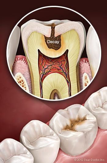
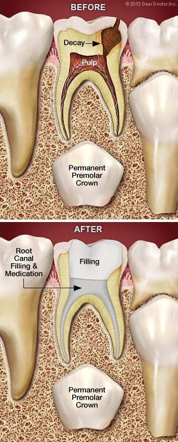
Unlike its hard outer surface, the soft pulp inside the tooth is rich in blood vessels and nerves. Problems in this area are often signaled by tooth sensitivity and pain. When these symptoms occur, radiographs (x-rays) are often necessary to confirm that the pulp is diseased, or dying. That’s when treatment is needed, before an abscess or further infection can develop.

In severe cases, the tooth may need to be removed, and a space maintainer installed to fill the gap. But many times, space maintainers don’t fully restore the tooth’s functionality. Plus, they are susceptible to coming loose and must be monitored constantly. If possible, other treatment methods are preferred, such as:

  • Indirect pulp treatment. If pulp damage is minimal, it’s possible to remove most of the decay (but not the pulp), apply an antibiotic, and then seal the tooth up again; that’s referred to as an “indirect” treatment.
  • Pulpotomy. Alternatively, if decay is limited to the upper portion of the pulp, we may recommend a “pulpotomy.” This involves removing the damaged part of the pulp, stabilizing the remaining healthy portion, and then disinfecting and sealing the tooth. This “partial” root canal is a time-tested technique that’s successful in many cases.
  • Pulpectomy. If pulp tissue is infected through the entire tooth structure, a pulpectomy may be needed, which requires the removal of all pulp tissue. The canals are then disinfected, shaped, then filled and sealed with inert material. Afterwards, the crown (visible part) of the tooth will be restored. This resembles traditional root canal therapy, with a crucial difference: The sealant we use in children is capable of being dissolved by the body. That way, when it’s time for a permanent tooth to erupt, the baby tooth’s roots can be naturally absorbed and tooth development can proceed normally.

Preparing for Your Child’s Root Canal Treatment

As you probably already know, most of the legends you may have heard about root canal therapy simply aren’t true. In fact, the procedure generally causes little discomfort, but is quite successful in relieving tooth pain! Dentists are adept at using anesthesia to block the sensation of pain, and are experienced in calming the fears of young ones. While it’s understandable that you may be nervous, it will help if you don’t let your child pick up on your own anxiety. A calming voice and a gentle touch can do much to relieve stress.

After a thorough examination, the best options for your child’s treatment will be recommended. These procedures are routine and follow-up instructions will be provided. A root canal is nothing to fear: Think of it as a treatment that may save your child from some tooth pain now, and potentially a lot of corrective dental work later on.

TMD

If you experience ongoing pain in the area near your ear, your jaw or the muscles on the side of your face, possibly accompanied by a clicking or popping sound or restricted jaw movement, you may be suffering from TMD — an abbreviation for Temporomandibular disorders. Sometimes people incorrectly use the term TMJ to refer to these problems, when in fact TMJ is the abbreviation for the temporomandibular joint — or jaw joint — itself. So while you definitely have a TMJ (two of them in fact), you may or may not have TMD.

TMD, then, describes a group of conditions characterized by pain and dysfunction of the TMJ and/or the muscles surrounding it. It’s not always so easy to figure out exactly what’s causing these symptoms, but the good news is that most TMD cases resolve themselves with the help of conservative remedies that you can try at home. In fact, it’s important to exhaust all such reversible remedies before moving on to anything irreversible, such as bridgework or surgery.

The two TMJs that connect your lower jaw, the mandible, to the temporal bone of the skull on either side, are actually very complex joints that allow movement in three dimensions. The lower jaw and temporal bone fit together as a ball and socket, with a cushioning disk in between. Large pairs of muscles in the cheeks and temples move the lower jaw. Any of these parts — the disk, the muscles or the joint itself — can become the source of a TMD problem. If you are in pain, or are having difficulty opening or closing your jaw, a thorough examination can help pinpoint the problem area; then an appropriate remedy can be recommended.

Causes of TMD

As with any other joint, the TMJ can be subject to orthopedic problems including inflammation, sore muscles, strained tendons and ligaments, and disk problems. TMD is also influenced by genes, gender (women appear to be more prone to it), and age. Physical and psychological stress can also be a factor. In some cases, jaw pain may be related to a more widespread, pain-inducing medical condition such as fibromyalgia (“fibro” – connective tissues; “myo” – muscle; “algia” – pain).

Signs and Symptoms of TMD

Clicking Sounds — Some people with TMD hear a clicking, popping or grating sound coming from the TMJ when opening or closing the mouth. This is usually caused by a shifting of the disk inside the joint. Someone standing next to you might even be able to hear it. Clicking by itself is actually not a significant symptom because one third of all people have jaw joints that click, studies show. However, if the clicking is accompanied by pain or limited jaw function — the jaw getting “stuck” in an open or closed position, for example — this would indicate TMD.

Muscle Pain — This can be felt in the cheeks (masseter muscles) and temples (temporalis muscles), where the two big pairs of jaw-closing muscles are located. If you feel soreness and stiffness upon waking up in the morning, it’s often related to habits such as clenching and/or grinding the teeth at night. If you have this type of nocturnal habit, a custom-made nightguard should be very helpful in decreasing the force applied to your teeth, which will in turn allow your muscles to relax and relieve pressure on your jaw joints. Other self-care remedies are discussed below (please see Relieving the Pain).

Joint Pain — Pain that’s actually coming from one or both jaw joints technically would be described as arthritis (“arth” – joint; “itis” – inflammation) of the TMJ. Radiographs (x-ray pictures) show that some people have arthritic-looking TMJs but no symptoms of pain or dysfunction; others have significant symptoms of pain and dysfunction but their joints look normal on radiographs. There is no cure for arthritis anywhere in the body, but medication can sometimes help relieve arthritic symptoms.

Relieving the Pain

Once you have been examined, a strategy for treating your condition and managing your pain can be developed. Sometimes a temporary change to a softer diet can reduce stress on the muscles and joints. Ice and/or moist heat can help relieve soreness and inflammation. Muscles in spasm can also be helped with gentle stretching exercises. Non-steroidal anti-inflammatory medications and muscle relaxants can also provide relief.

Other Treatment Options

Severe TMD cases may require more complex forms of treatment, which might include orthodontics, dental restorations like bridgework, or minor procedures inside the joint such as cortisone injections or lavage (flushing) of the joint. It’s rare for major surgery ever to be necessary in a case of TMD. Again, it’s important to try the wide range of conservative, reversible treatments available, and give them enough time to work as they almost always prove effective. The first step is an examination at the dental office. To learn more about available treatment options, view this Chart on TMD Therapy

Intra-Oral Camera

The intra-oral camera is a valuable tool dental professionals can use to help you understand your examination, diagnosis and treatment. This small, handheld video camera is about the same size as a dental mirror (or an oversized pen) and comes with a disposable plastic sheath for contamination prevention. It is used to take actual pictures of your teeth with up to 25 times magnification and project them onto a screen for your review. It can also be used to give you a video tour of your entire mouth so that you can see things such as plaque deposits, decay, worn teeth, and broken or missing fillings. Lastly, pertinent images can be printed for your patient file for future reference — or even for you to take home.

A Picture Is Worth a Thousand Words

This adage is proven true many times each day in offices equipped with an intra-oral camera. Prior to the development of this technology, some patients found it challenging to understand problems such as dental decay and periodontal disease. Now, it’s possible to display, pause, and zoom in during a video examination of the problem area, so that you can see it for yourself — all in color and crystal clear.

Advantages of an Intra-Oral Camera

  • An intra-oral camera makes you more of an active partner in your dental treatment, enabling you to see what a dentist sees — an especially important benefit when additional treatments need to be discussed, or when treatment options must be explained — so that you can make informed decisions.
  • You may be more comfortable asking questions and better able to understand a treatment option or oral hygiene concern when you can actually see it.
  • Because it gives you the real-world picture of your dental hygiene, the intra-oral camera is ideally suited to show you techniques for improving your oral health and hygiene habits.
  • With its powerful magnification (much superior to the naked eye), it reveals the early stages of maladies such as gum disease and cavities.
  • The images can be captured and reexamined later, to show you changes in your oral health and hygiene or how a multi-phase treatment is progressing.
  • Furthermore, it can provide insurance companies with the proof they require to approve a needed treatment.

Tooth Decay Prevention

Tooth Decay.

Tooth decay is often called the second most prevalent human disease, after the common cold. Without effective treatment (as was the case through most of history) it can lead to pain, tooth loss, and sometimes worse illnesses. Even today, it’s estimated to affect over a quarter of U.S children from ages two to five, and half of those aged 12-15. But it doesn’t necessarily have to! You can take steps to prevent tooth decay from harming your teeth — or those of your loved ones.

There’s one important fact you should understand up front: No single “magic bullet” can stop tooth decay in every case. Instead, fighting decay should be viewed as a process of preventive maintenance, like taking care of your car — except that (unlike a car) your natural teeth, with proper care, can last a whole lifetime. The basic aspects of this process are practicing good oral hygiene at home, and coming in to the dental office for regular cleanings and checkups.

If you’ve been in the dental office for routine visits, you’re probably already familiar with the special tools dentists use to remove buildups of plaque (a bacterial biofilm) and tartar (a hardened deposit, also called calculus) from your teeth. Hand-held instruments, ultrasonic scalers, or both may be used to give your teeth a thorough cleaning. Afterwards, your teeth are thoroughly checked for decay, and cavities are treated when necessary.

Yet there’s still more that can be done to prevent tooth decay. Could your diet be a contributing factor? Is your brushing technique adequate? Could you benefit from additional preventive treatments? Today, with our increased understanding of what causes tooth decay and how to treat it, it is possible to focus on what decay prevention tactics would work best in your particular case. In fact, it’s now possible to assess each individual’s risk factors for decay, and concentrate on doing what’s most effective for you.

How Does Decay Start?

Tooth Caries Balance.

It’s useful to think of the mouth as a dynamically balanced ecosystem, in which living organisms, including helpful and harmful bacteria, are constantly interacting. When conditions are right — namely, in the presence of certain sugars — some pathogenic (harmful) bacteria produce acids that cause teeth to lose minerals and begin breaking down. Even a diet having excessive acidic foods can influence deminerialization of your teeth. But in more favorable conditions, the damage these pathogens do is undone by the body’s own healing mechanisms — which includes your healthy saliva.

A major goal in decay prevention is to tip the balance in favor of the beneficial processes. Keeping up a regular habit of brushing and flossing, getting adequate fluoride, and a diet with limited acidic foods is certainly helpful. Yet even with these measures, some individuals will be more prone to tooth decay than others, and may need extra help and guidance.

Additional Steps to Prevent Tooth Decay

If you’re one of these individuals, it may help you to learn effective brushing techniques and practice other measures at home — for example, using special toothpastes or mouthrinses. When necessary, in-office treatments such as topical fluoride applications are available. If you aren’t getting enough fluoride through drinking water or other sources, this treatment can help prevent tooth decay. Anti-bacterial treatments may also be beneficial in some cases, as is nutritional counseling.

Dental Sealants.
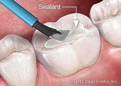
Finally, if your child’s teeth are susceptible to tooth decay, consider having a dental sealant applied. This is a practically invisible layer of plastic resin that is placed on the top (chewing) surfaces of the back teeth. It’s a painless procedure that fills in the natural pits and folds of the tooth, making them much more resistant to bacterial damage.

So, don’t think that tooth decay is inevitable — instead, find out what you can do to help prevent this disease from affecting you or your loved ones.

Bone Grafting

Do you think of bone as a hard, rigid material that never changes? In fact bone remodels itself all the time: Your body is constantly depositing new bone cells and removing old ones. In the case of the bone that supports your teeth, this process can be helpful or harmful. For example, the jawbone’s adaptability allows an orthodontist to move teeth into a better position with braces. But in the case of losing a tooth as an adult, the bone changes that result can have serious consequences.

When teeth are lost, the bone that used to surround them begins to melt away or “resorb.” Tooth-supporting bone can also be lost when you have periodontal (gum) disease. If you loose enough teeth and bone, your facial features will sag, giving you a more aged appearance; it can also complicate treatment to replace your missing teeth. Fortunately, with modern bone grafting-techniques, the bone that has been lost can be built up again. This can benefit both your health and appearance by strengthening your jawbone, allowing more effective tooth replacement, and increasing support to your facial features.

Bone grafting is a minor surgical procedure that is normally done in a dental office. An incision is made in your gum to gain access to the bone beneath it, and then grafting material is added. Most often, the grafting material is processed bone minerals around which your body will actually deposit new bone cells.

The grafting material itself can come from your own body, but very often it is bone from an animal or human donor that has been treated by a laboratory to make it sterile and safe. It can even be a synthetic substance. Grafting material comes in a variety of forms: powder, granules, putty or a gel that can be injected through a syringe. The graft, which is generally covered by a collagen membrane for optimum bone repair, will act as a scaffold onto which your body will build new bone.

Uses for Bone Grafts

Bone grafting for dental implants.

Bone grafts are used in dentistry to accomplish the following treatment goals:

  • Saving Teeth — When severe periodontal disease causes bone loss, teeth can become loose and at risk of being lost. In order to save them, the bone around them can be regenerated through grafting; this increases bone support and helps keep them in place.
  • Tooth Extractions — These days, it is very common to deposit bone grafting material into a tooth socket after a tooth has been removed. That way, should you want to replace your tooth with a dental implant later on, that option will be available.
  • Dental Implants — In this optimal tooth-replacement system, a small titanium post embedded in the jawbone is attached to a highly realistic dental crown, permanently replacing the missing tooth. Implants require good bone volume and density to achieve their excellent functionality and high success rates. If you have already experienced bone loss, a graft can help regenerate enough bone to place the implant successfully.

What to Expect

The procedure for placing a bone graft usually requires only local anesthesia, though oral or IV (intravenous) sedatives can also be used to achieve a higher state of relaxation. Since a small incision is made in your gum tissue to gain access to the bone that will receive the graft, you may experience some soreness in the area after the surgery. This can usually be managed by over-the-counter anti-inflammatory medication and/or pain relievers, as well as ice therapy after the procedure. Any discomfort should only last a day or two. Then, over the next several months, your body will replace the graft with its own bone, reversing the decline in bone quantity you have experienced.